
3. Cloning of Gastrointestinal Villi in Cell Culture from Single, Isolated Cells
Normal human living intestine was obtained from surgical operations, and enzymatically digested so as to obtain dispersed single cells from the intestine samples. The suspended intestinal cells were cultured in MEM medium and divided into two groups: test group and control group. The MEBO regenerative nutrients were added to the cultures in the test group, while the control group did not receive any supplement. Every 3 days the old growth medium was discarded and fresh growth medium same as the first round was added to the cultures in the respective groups.
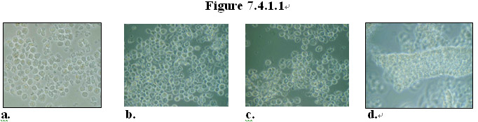
After 55 days of culture, in the treatment group the cells had grown into groups of cells appearing in various forms, including proliferation and differentiation of the cells isolated from human intestine samples. As shown in Figure 7.4.1.1a-c, in some areas, some of cells formed clusters that appeared to look like pieces of tissue. Surprisingly, it was observed that in some areas, tissue pieces resembling a fully assembled villus structure appeared in the culture Figure 7.4.1.1d. In comparison, most cells in the control group had died by Day 55.
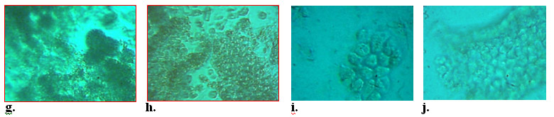
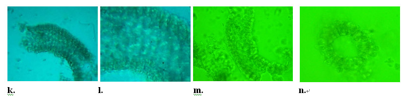
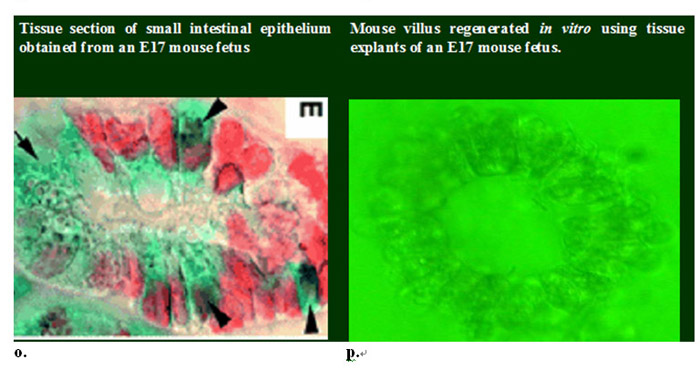
To verify whether intestinal villus can be formed from proliferation and differentiation of isolated dispersed intestinal cells in vitro in the presence of the MEBO regenerative nutrients, experiments were conducted using intestines isolated from a mouse fetus (Figure 7.4.1.2a-c). The mouse fetal intestine was cut into small explants and cultured in cell culture medium in the presence or absence (control) of the MEBO regenerative nutrients (Figure 7.4.1.2d-f). Consistent with observation of cells from human intestines, single cells migrated from the explants started to form clones (Figure 7.4.1.2h-i) in the culture which grew to form tissue pieces having the same structure of a villus were formed in the presence of the MEBO regenerative nutrients (Figure 7.4.1.2j-m). In comparison, intestine explants in the control culture disintegrated (Figure 7.4.1.2g). A cross-sectional view (Figure 7.4.1.2n, p) of the villus shows that the tissue formed in vitro in the presence of the MEBO nutrients has the same structure as that obtained from tissue sections of intestines of a mouse fetus (Figure 7.4.1.2o) as shown in Yang et al. (2001) “Requirement of Math1 for secretory cell lineage commitment in the mouse intestine” Science 294:2155-2158.
These results are groundbreaking as they demonstrate that the MEBO regenerative nutrients can sustain vigorous proliferation and facilitate orderly differentiation of somatic cells for a long period of time to form multiple cell types that are linked together to form tissue with physiologically correct structure in the culture. From single, isolated cells from human and mouse intestines at least 4 types of cells are formed: entercytes, mucus-secreting goblet cells, enteroendocrine cells, and Paneth cells which are originated from epithelial stem cells in the crypts of the intestine. Although it remains to be ascertained the nature of the single, isolated cells that produced the multiple cells types in the culture containing the MEBO nutrients, it is significant that physiologically correct tissue has been cloned in vitro from single cells isolated from intestines. To our best knowledge, there is no report from others with the same results.

 【HBRRS: Human Body Regenerative Restoration Science】
Human Body Regenerative Restoration Science will bring about four revolutions to human world: the scientific revolution of human life, agriculture industrial revolution, food industrial revolution, and life energy revolution, which will lead us entering the brand new human regenerative life world - XU Rongxiang